

Two innovative vascular phantoms from the MANTA research project enabled doctors at the South and Central German Radiology Congress in Hof to practice complex medical procedures. These artificial replicas of human blood vessels simulate important anatomical and physical characteristics of the body and allow for the training of medical treatment techniques. The university itself received valuable feedback from the presentation that will aid in the further development of its models.
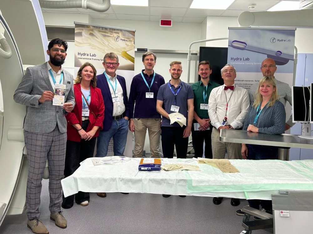
How can complex medical procedures be practiced without relying on patients? A research project at Hof University of Applied Sciences has been addressing this question since the middle of last year, and its findings were recently put to use at the South and Central German Radiology Congress in Hof. Two training models from the registered HYDRA LAB brand were made available to participants for hands-on exercises. Over the course of three days at the congress, radiologists were able to use them to practice various interventional radiology techniques under realistic conditions and under supervision.
Practicing medical procedures without endangering patients
The focus is on a procedure known as embolization. In this minimally invasive procedure, radiologists insert a thin catheter through a small access point—usually in the groin or at the wrist—and guide it through the blood vessels to a specific location in the body. There, they can, for example, selectively seal off blood vessels to stop internal bleeding. Such a procedure can be lifesaving, especially in cases of acute abdominal bleeding. However, the necessary techniques and the safe handling of the instruments require a great deal of experience and practice.
This is exactly where the training models from Hof University of Applied Sciences come in: These so-called phantoms technically replicate the human vascular system, making it possible to practice medical procedures outside the operating room. The models feature a transparent, flexible vascular system through which a pump circulates fluid at adjustable pressure and flow rates. This allows for the simulation of key characteristics of the human circulatory system.
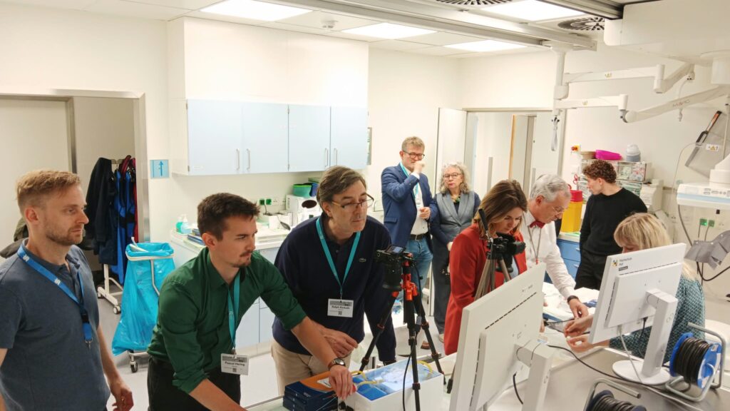
Doctors can navigate through the simulated vessels using special microcatheters and guidewires and employ various materials to occlude blood vessels. These include, for example, so-called coils—fine metal spirals that are precisely placed within a vessel—as well as special tissue adhesives. This allows various treatment techniques to be repeatedly tested and practiced without exposing a patient to risk.
Cost-effective technology with international potential
The models are being developed as part of the MANTA (Medical Angiography Simulation Phantom for Training Prospective Physicians) research project at Hof University of Applied Sciences. The project leader is Prof. Dr.-Ing. Anke Müller, who is also dean of the university’s Faculty of Engineering and Applied Design. René Göhring is responsible for the technical implementation of the training models at Hof University’s MakerSpace, while Prof. Thomas Rausch oversees the design of the simulated vascular system, which is manufactured by lab staff members Nathalie Koch and Martin Thoß.
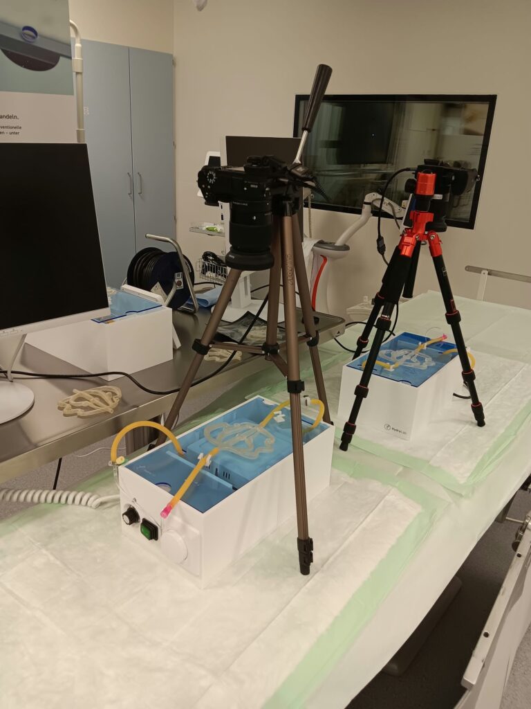
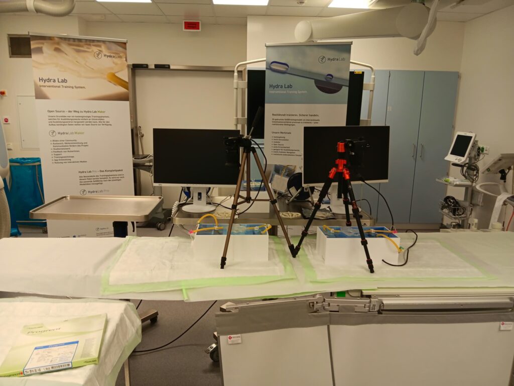
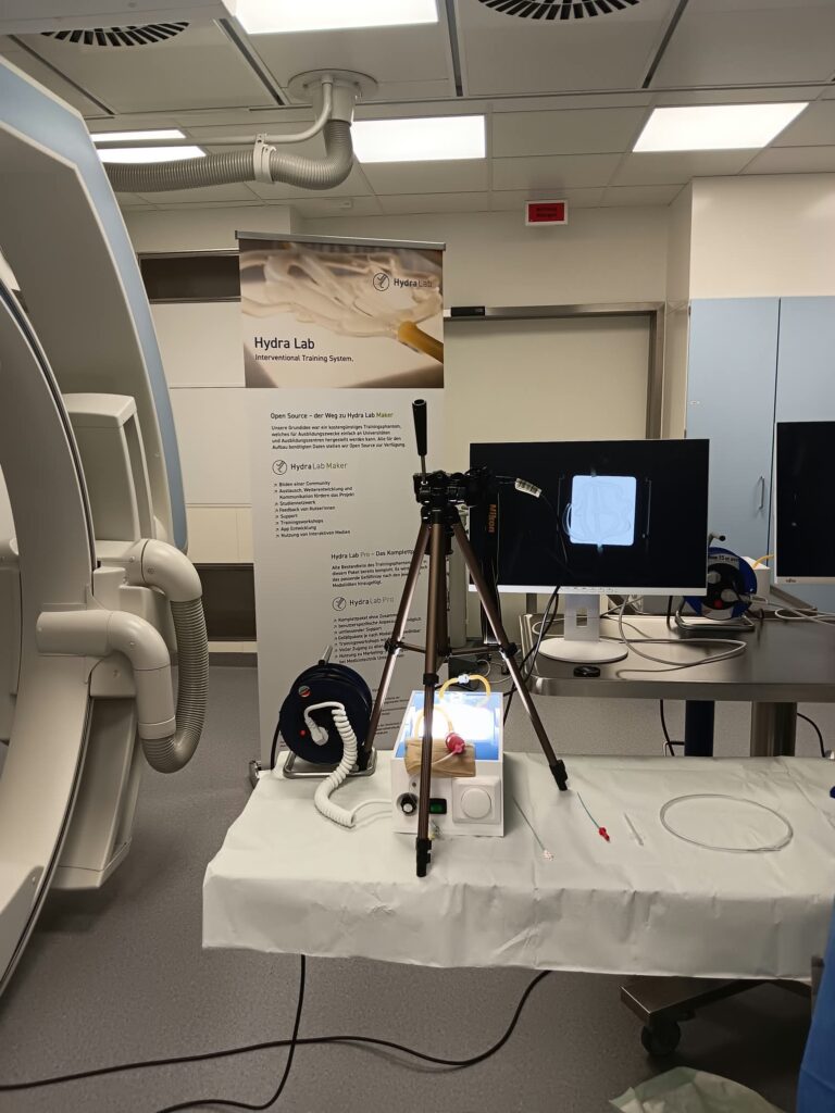
A unique feature of the project is its approach to combining a simulation that is as realistic as possible with manageable manufacturing costs. The models are created using additive manufacturing processes—that is, 3D printing—and off-the-shelf components. This sets the development apart from many commercial medical training systems, which involve significantly higher acquisition costs.
“Our goal is to develop a training system that simulates complex medical procedures as realistically as possible while also being cost-effective and easy to manufacture, so that it can be made available to as many medical facilities as possible—including those in other countries. ” “Doctors should have the opportunity to practice important procedures and various treatment techniques under controlled conditions as often as necessary for their safety and to build routine. In the long term, this can also improve the quality of patient care.”
Prof. Dr.-Ing. Anke Müller, Project Leader
In the long term, the project also pursues an open-source approach. The basic idea is to make the design and development principles accessible so that the models can be replicated at other locations and adapted for additional medical applications. Possible variations include models for other organ systems, for urology, or for patients of different sizes. This could open up new possibilities, particularly in countries where expensive medical training systems are scarce.

Hands-on testing under the guidance of experienced radiologists
At the radiology conference in Hof, the training models were made available for the first time to a broader group of medical professionals in a comprehensive hands-on training format. Under the guidance of course instructors Prof. Dr. Boris Radeleff and Prof. Ralph Kickuth, participants tested various embolization techniques. For the research team at Hof University of Applied Sciences, this direct contact with medical practice was particularly valuable. After all, it is only when the models are used by experienced radiologists that it becomes clear how well they can be integrated into specific training procedures, which features are already convincing, and where there is still room for improvement.
“Feedback from the medical field is extremely important for our future work. When experienced radiologists use our models, we receive immediate insight into how realistically certain procedures can be simulated and where we can still make technical improvements. It is precisely this exchange between the university and the medical field that drives our development forward.”
Prof. Dr.-Ing. Anke Müller
Next Appearance at the German Radiology Congress in Leipzig
The next major presentation is already planned: In May 2027, the training models are also scheduled to be presented at the German Radiology Congress in Leipzig. In doing so, Hof University continues its collaboration with the medical community and is gradually bringing an innovative development from its MakerSpace closer to widespread use in medical education and continuing professional development.
The MANTA project is funded by the Hans Viessmann Technology Foundation and the Manfred Schlager Foundation.